Return To Book 3 Table Of Contents
The Fascinating Cerebellum… The Role Of Nitric Oxide… The Basal Ganglia…
Major Implications For Safety…
The cerebellum was said to be involved in coordination of both motor functions and cognitive functions - in the organization of our thoughts and motions! How interesting – again.
Fragmented thought, a “hallmark” of autism, schizophrenia, and Alzheimer’s and the clearly impacted part of the brain in autism – the cerebellum – known to be associated with the “organization of thoughts”! Surely, understanding the cerebellum would be key!
The cerebellum was also believed to be that part of the brain that "changed" the most during adolescence. Again, how very interesting. From reading parent discussion boards, it seemed to me that many parents complained their children with autism seemed to “get worse” at the onset of puberty and it was a known fact that children with autism were known to develop seizures in adolescence. Although some studies indicated that approximately thirty percent of children with autism developed seizures, I suspected that in actuality, there were many more than thirty percent. The simple fact was that most parents did not realize “blank stares”, “purposeless motion”, “picking at things” were all signs of seizure activity – and all things found in autism!
In my second book, Breaking The Code To Remove The Shackles Of Autism: When The Parts Are Not Understood And The Whole Is Lost! I had commented that “blank stares” appeared to be a coping mechanism in children with autism. I still believed that to be true – to an extent. However, the more I learned about autism, the more I came to see that perhaps blank stares in children with autism were more a sign of a seizure.
We all had those “blank stares” when we were thinking or retrieving information – and that, I did believe to be very true in children with autism, children whom I truly believed “lived via reference” and hence were constantly engaged in “information retrieval” – which could certainly account for “blank stares”, but now, I saw that these could truly involve much more than “information retrieval” – that “blank stares” could certainly be signs of seizures!
As with everything in matters relating to autism – I, too – was learning and at times, that obviously meant looking at things in a new light when new information presented itself. As such, I always reminded families that in reading any of my materials – look for what “made sense” – and “pitch” the rest. I was simply sharing my thoughts and opinions – in case they helped others with “their puzzle” too, and obviously, as I learned more, my thoughts or opinions on certain issues could potentially change over time. I had no problem with admitting that. This was certainly not an issue of pride for me – it was one of attempting to get to the truth – of attempting to understand my son – in order to best help my son!
As I had done on so many occasions, I began to dig much deeper into many issues and was amazed at how quickly so much now seemed to fall into place. Perhaps one of the best sources of information was an interview by PBS’s Frontline with Dr. Jay Geidd, neuroscientist and chief of brain imaging at the National Institutes of Mental Health (NIMH) called: Inside The Teenage Brain. Although this interview had nothing to do with autism, it certainly provided valuable insights into the understanding of the cerebellum! In this interview, Dr. Geidd stated – and I quote -
"Identical twins' cerebellum are no more alike than non-identical twins".
Thus, identical twins, twins who had come from one hundred percent the same genetic code, the same cell, had cerebellums that were no more alike that non-identical twins or persons who had come from different cells to start with. Dr. Geidd further stated that we knew the cerebellum continued to grow and mature at least until the early twenties. Because of this, Dr. Geidd further stated he believed the cerebellum to be impacted more by environmental factors than genetics. Environmental factors certainly could include mercury, aluminum or iron poisoning or exposure to viruses. In this interview, Dr. Giedd stated that the cerebellum was “not very genetically controlled”. Again, how interesting! The “peak” for diagnosing schizophrenia in males was in the early twenties. In females, schizophrenia was usually diagnosed in the late twenties. All this seemed to very much argue against a “genetic link” for autism – and possibly for schizophrenia.
We had researched the "genetic link" to autism for over sixty years and the "genetic link" to schizophrenia and Alzheimer’s both for close to one hundred years. Perhaps it was truly time to start looking elsewhere – to environmental factors – like mercury, aluminum and/or iron poisoning and the impact of viruses in vaccines.
Insanity is when we keep doing the same thing and keep expecting different results.
Albert Einstein
If this was how one of mankind’s most brilliant minds had defined insanity, I suspected there existed a little “insanity” within the very walls of the National Institutes of Mental Health! Albert Einstein was indeed a brilliant man – a man with the ability to describe both - the behavior and the prognosis – using the same word. “Insanity” after all was how many in society had for so long described “schizophrenia” – a disorder I now so painfully saw, had been so very, very misunderstood by society – a disorder I now saw as but - another on the life spectrum - that truly involved so many disorders.
Confusion certainly did seem to reign everywhere when it came to mental illness. On the one hand, we had a neuroscientist and the chief of brain imaging at the National Institutes of Mental Health (NIMH) telling us that the cerebellum was controlled not by “genetics” but rather more by “environmental factors” and yet, it appeared that in matters relating specifically to the study of autism, a disorder known to have serious implications in terms of the cerebellum, at the same National Institute of Mental Health, when studying autism specifically, we seemed to assume only "genetics" or problems in pregnancy played a role in why the cerebellum was so impacted in autism! Indeed, at the National Institutes of Mental Health, we seemed to refuse to want to investigate or fund any studies that might show a link between autism and vaccines.
Why was it that given the public outcry as it related to increasing rates of autism that the National Institutes of Mental Health continued to fail to investigate the issue of a possible link to mercury poisoning or vaccines in general? Was this organization not funded by public funds? If so many were pointing the finger to vaccines - why the lack of any independent study and significant funding to investigate this issue! The public should allow no excuse of “no money in the budget for such studies at this time”. This government could find billions – overnight – if it had to. Had there not been enough warning bells, sounding, for the CDC, NIMH and the NIH? I suspected we all knew that the answer to that question was because the CDC, NIMH and NIH knew the public's suspicions may very well be proven correct and so, everything but vaccines would be looked at as the public was told by the government that it was busily "looking for answers".
Yes, there had been many, many studies into autism, but, quite frankly, many of them were so utterly ridiculous that I could barely believe public funds were being used for some of this “science”. For example, there had been studies trying to link “lack of sunlight” to autism. In my opinion, any “findings” from such studies would indeed have a very difficult time explaining the explosion in autism in places like California and Australia. Although, given that sunlight was tied to the breakdown of bilirubin, perhaps those scientists wondering about the role of sunlight should be looking not simply at sunlight itself but at the role of sunlight as it related to a possible bilirubin and autism link!
When was the public going to finally demand honest answers in matters relating to the possible autism-vaccine connection? Would the fact that Alzheimer’s and schizophrenia now appeared to play into this be enough for public outcry to finally be heard? Would the fact that so many of us were heading for Alzheimer’s – with fifty percent impacted over age eighty five – be enough to finally “motivate” society into demanding honest research and honest answers into these issues?
You would think that in over eighty years, a study investigating the safety of mercury in vaccines would have been conducted at least once - but according to Congressional Hearings in June of 2002 - that was not the case - there had not been even one study by the government on the safety of mercury in vaccines! Perhaps it was time to start looking into what parents of children with autism considered the most obvious place to look – the autism-vaccine link! Truly, with no study on the safety of mercury in vaccines in over eighty years, and increasingly aggressive vaccinations schedules, the government had been asleep at the switch – for decades. No study on mercury – and vaccine studies that lasted only a few days to a few weeks! Personally, my comfort level in terms of what we knew of the safety of vaccines had gone completely out the window!
Yet, even with no studies into the safety of mercury or long term studies into the safety of vaccines in general, there were other studies – studies not related to autism or vaccines - but studies from which a great deal could be learned and studies that were – still – relevant to autism, schizophrenia and Alzheimer’s. It would be within those studies that I continued to search for answers.
Scientific research into the functions of the cerebellum showed the cerebellum was known to control the muscles that were used in speech! Given that the cerebellum had, clearly, been implicated in autism, was it any wonder that up to fifty percent of children with autism were considered non-verbal! But, what else did the cerebellum do?
The cerebellum, in the past, had always been viewed as primarily a coordinator of motor functions. As such, it was – for a long time - thought to be a more “primitive” part of the brain. But, even an amoeba had "motor" functions. And - even a squirrel - could twirl a nut while balancing himself on a twig! How was it that even the most primitive creatures could have fine motor functions and yet, in humans, it took more than twenty years for the cerebellum to develop - that part of the brain most closely associated with the coordination of motor functions in humans appeared to take the longest of all to mature! There had to be a great deal more to the cerebellum than simply motor coordination.
I soon discovered the cerebellum indeed played a major role in much more than just the coordination of motions. It was also involved in the coordination of “higher thoughts” language and emotions. The cerebellum, in animals, was also known to be associated with the tracking of moving objects – and the same appeared to be true in humans. As more and more was learned about the cerebellum, it became evident the cerebellum was involved in the regulation of sensory data. Again, this was all very intriguing to me since I knew that my own son had great difficulty perceiving moving vehicles.
Indeed, the cerebellum was now being compared to a mini super computer in charge of various "regulating" or “coordinating” functions.
If the cerebellum was involved in the "regulation" of things, could it not be involved in the “need for sameness” in things. Motor activity and higher thought processes were known to occur in the frontal lobe. Damage to the frontal lobe resulted in obsessive-compulsive behavior. If the cerebellum was involved in the coordination of motor functions, and the coordination of “higher thoughts”, would damage to the cerebellum not also result in obsessive thoughts (i.e., the need for sameness in everything), leading to obsessive thoughts and behaviors or obsessive thoughts and obsessive motor functions?
The more I studied the cerebellum, the more I became convinced that this “regulator” and “coordinator” could indeed be the “brains of the brain”. Of course, that was simply “my theory”. As science moved forward, I would not, however, be surprised to see science discover the cerebellum to be the overall body regulator – of thoughts – of motions – and perhaps regulating everything from basic motor functions to life centers in the brain stem, as well as possibly regulating the immune system function and other functions throughout the body.
Of course, that was just "my theory” at this point as to the possible critical role of the cerebellum, but it certainly would put a great many pieces of the puzzle into place. For example, if the cerebellum indeed was the “regulator” of life functions, then, it would make perfect sense that it be located near the brain stem – in order to more quickly regulate “life functions” or “coordinate” life functions in the brain stem with say motor functions in the frontal lobe. We knew that respiration was in the brain stem. But, could the cerebellum play some role in actually controlling respiration as it related to current motor activities?
The more I read, the more my suspicions about the role of the cerebellum seemed to be confirmed. For example, research had found that the immune system talked to the brain via the blood. Nitric oxide was a gas-like neurotransmitter known to play a role in the flow of blood. Indeed, it appeared that of all brain areas, the cerebellum had the most to do with nitric oxide levels. Nitric oxide was synthesized as needed by NO synthase (NOS) from its precursor L-arginine. Although I did not have a background in science to “fully appreciate” the meaning of that, the fact remained that NOS seemed to be in rather high concentration in – the cerebellum – and that was something I could understand. If high concentrations of (NOS) were found in the cerebellum, it stood to reason that nitric oxide would be produced in high concentration where (NOS) was found – and that would be - in the cerebellum!
Nitric oxide was believed to increase the permeability of the blood brain barrier. … the cerebellum… the immature blood brain barrier of an infant… the cerebellum… a “regulator”… could the cerebellum somehow be involved in regulating the maturity of the blood brain barrier too? Could the cerebellum be involved in regulating the permeability of the blood brain barrier? Increasing permeability was one thing – actually regulating it was quite another! Yet, somehow, it certainly would seem to make sense – at least in my opinion – but again, this was just “a theory” I had at this point.
Although nitric oxide was considered a "neurotransmitter" – outside the body – it was considered a toxin. Within the body, it was known that too much nitric oxide could lead to cell death! Was the cerebellum in autism, schizophrenia and Alzheimer’s producing too much nitric oxide? Was it really a neurotransmitter or was it a neurotoxin that had been implicated in “altering” many functions within the brain/body? Was this a neurotransmitter – or a neurotoxin? Could it be both? Could it be just a matter of “the level of nitric oxide”? Given nitric oxide was said to be “unlike” any other neurotransmitter previously known to man – because nitric oxide was a gas - I could not help but ask the obvious question: Were we really sure this stuff belonged in the body in the first place?
I knew that scientists out there would have the initial response of “well, that’s an absurd question – of course, it is a neurotransmitter”, yet, given it was “so different” from other neurotransmitters, was it not possible that this could be a neurotoxin with many implications if found within the body – some perhaps good – but others, potentially very bad?
When I thought of nitric oxide and the whole “toxin if found outside the body thing”, it was just another one of those “things” that for me – was difficult to come to grips with and as such, I had to ask the question – as crazy as surely, I knew it had sounded to so many. This was a “simple” question – “simple” could mean “simple minded”, but if my question was “that crazy”, then, surely, it would be easy to answer. Surely, there was a reason as to why this substance could be perhaps “good”, but perhaps very devastating also.
How was it that a toxin outside the body could be good within the body? As had been the case with “breastmilk jaundice” and so many other issues, I just had a really, really difficult time with that issue. Perhaps it truly was “a neurotransmitter” – but what if it was really a toxin that did not belong there! This gas-like neurotransmitter it seemed, had only recently been identified – the question was – did it really belong there? Certainly, man made new discoveries relating to the brain and its functioning each day. Thus, yes, absolutely it was possible that this was indeed a “neurotransmitter gas”. My intent here had simply been “to raise a question” – because it appeared to me that there seemed to be tremendous “negatives” associated with nitric oxide, too!
If nitric oxide was a neurotransmitter gas and it did belong in the body, then I had another obvious question: What was leading to abnormal levels of nitric oxide production – known to exist in Alzheimer’s, schizophrenia and autism. If nitric oxide was most closely associated with (NOS) and (NOS) was found in rather high concentration in the cerebellum, and nitric oxide was also associated with cell death, could the damage to the cerebellum in children with autism not have resulted from excessive levels of nitric oxide? Could mercury or aluminum have caused excessive levels of nitric oxide? Could viruses have caused excessive levels of nitric oxide? Nitric oxide was known to bind to dna and have a role in immune system functions.
As I researched – I found what appeared to be another piece to the puzzle. Nitric oxide was known to bind with iron! Could excess iron – iron overload - have led to excess levels of nitric oxide?
The fact that nitric oxide was known to bind with iron and the fact that persons with autism and Alzheimer’s were often known to suffer from iron overload, truly made me wonder – again – as to the role of iron in all this! Iron, I knew, was a substance that could bind to many, many things. Yet, it certainly seemed to “come up” in critical “trouble areas” when it came to these disorders. In addition, it was a medically and scientifically proven fact that males retained more iron than women. Could the iron connection possibly contribute to the higher and earlier incidence of so many mental disorders in men - in boys? Could this explain why autism, generally, did not appear early on but rather only after a few years? How did iron metabolism change over time – specifically – from pre-birth on - in boys – in girls?
Excess iron was also known to suppress the immune system. Yet, iron was found in iron fortified formulas and baby foods - usually introduced around six months of age. Iron in breastmilk, was not believed to be influenced by the mother’s iron levels. But, excess iron in the mother during gestation could be passed to the unborn child.
Lactoferrin… nitric oxide … iron… iron… iron… so much seemed to point back to implicating an iron metabolism dysfunction – and liver dysfunction! Nitric oxide … known to bind to iron… could this be another means by which the immature infant’s system attempted to rid itself of excess iron given that the liver was not yet able to properly process iron?
High lactoferrin in spinal fluid in Alzheimer’s… a possible immune system response… low lactoferrin in autism… lactoferrin produced in the liver… an immature liver in infants... bile – not produced until six months of age - the only other source of lactoferrin other than breastmilk… lactoferrin binding to iron… and now… nitric oxide binding to iron, too – and (NOS) found in high concentrations in – the cerebellum – that part of the brain that appeared most impacted in autism!
Given Alzheimer’s was associated with “later stages of life”, a person afflicted by Alzheimer’s would have had a developed cerebellum, thereby, making that person potentially less susceptible to damage in the cerebellum. The fact that iron was not being properly processed as evidenced by iron overload in Alzheimer’s also indicated that the liver was dysfunctional.
In the case of an infant or young child exposed to excess iron or mercury – a substance known to interfere with the body’s ability to rid itself of iron – the cerebellum certainly could be impacted as nitric oxide synthase was found in highest concentration in this part of the brain and thus, provided “something” for iron to bind to in the absence of lactoferrin or proper liver function to process excess iron. Persons with schizophrenia, in my opinion, would have been “in the middle” in terms of cerebellum damage when considering brain developmental stages and liver function.
It certainly would be interesting to have a chart showing the “order of preference” in terms of “what” iron liked to bind to in the human body. My guess would be that, like lactoferrin, nitric oxide would be fairly “up there” on such a chart.
Interestingly, based on notes from the Simpsonwood meeting of 2000, a “behind closed doors” meeting attended by persons from the NIH, CDC, pharmaceutical industry, WHO, medical community, etc., it was a known fact that the earlier the exposure to mercury, the worse the effect on the person. In other words, exposure at one day was worse than exposure at ten days, was worse than exposure at one year, was worse than exposure at age twenty or age eighty. Persons attending this meeting had clearly indicated that they knew the earlier the exposure to mercury, the worse the outcome! Again, this was all very interesting indeed.
The following were a few quotes taken a report obtained by the US Autism Ambassador, LD Wedewer as they related to actual meeting notes from “the Simpsonwood meeting of 2000”:
Comments Per the Simpsonwood report were as follows:
“Dr. Keller, pgs. 116 & 118: "we know the developing neurologic system is more sensitive than one that is fully developed". [end of quote, emphasis added, CDC’s National Immunization Program (NIP) Report entitled Scientific Review Of Vaccine Safety Datalink Information, produced based on information from a June 7-8, 2000 meeting convened by CDC’s NIP Director, Dr. Walter Orenstein].
Dr. Verstraeten, pg. 162: "When I saw this, and I went back through the literature, I was actually stunned by what I saw because I thought it is plausible. First of all there is the Faeroe study, which I think people have dismissed too easily, and there is a new article in the same Journal that was presented here, the Journal of Pediatrics, where they have looked at PCB. They have looked at other contaminants in seafood and they have adjusted for that, and still mercury comes out. That is one point. Another point is that in many of the studies with animals, it turned out that there is quite a different result depending on the dose of mercury. Depending on the route of exposure and depending on the age at which the animals, it turned out that there is quite a different result depending on the dose of mercury. Depending on the route of exposure and depending on the age at which the animals were exposed. Now, I don't know how much you can extrapolate that from animals to humans, but that tells me mercury at one month of age is not the same as mercury at three months, at 12 months, prenatal mercury, later mercury. There is a whole range of plausible outcomes from mercury. On top of that, I think that we cannot so easily compare the U.S. population to Faeroe or Seychelles populations. We have different mean levels of exposure. We are comparing high to high I the Seychelles, high to high in the Faeroe and low to low in the U.S., so I am not sure how easily you can transpose one finding to another one. So basically to me that leaves all the options open, and that means I can not exclude such a possible effect." [end of quote, emphasis added, CDC’s National Immunization Program (NIP) Report entitled Scientific Review Of Vaccine Safety Datalink Information, produced based on information from a June 7-8, 2000 meeting convened by CDC’s NIP Director, Dr. Walter Orenstein].
To read more quotes from the "not for the public" Simpsonwood meeting 400 page report obtained by the US Autism Ambassador, readers were encouraged to go to the following link and “scroll down” to see the section on Simpsonwood:
http://autismawakeninginia.bizland.com/autismworldnewspaper/id47.html
I encouraged all families impacted by mental illness to at least make a copy of this valuable report – even if only for future reference.
This report had been made available for downloading by families around the world in the US Autism Ambassador's newsletter dated December 10th, 2002. This report was provided - in full - to Congressman Dan Burton by the US Autism Ambassador as official testimony submitted on behalf of the public for the December 10th 2002 hearings relating to vaccine issues and government reform in Washington! Also, provided, as official testimony submitted on behalf of the public for the December 10th, 2002 hearings was another report – one relating not to mercury – but to aluminum. This report – another report of several hundred pages - had resulted from the “Puerto Rico meeting of 2000 on aluminum”. In this report, attendees seemed to indicate that aluminum was as much a concern as mercury! Very eye opening comments indeed! Several key Democrats and Republicans had also been provided with this information – at both federal and state level!
Thus, the more immature the cells when exposed to mercury – the worse the impact. Given this observation, could we not infer something else – that perhaps the most immature of the immature cells would be the most susceptible. Since the cerebellum was known to take twenty years to mature, that certainly would make cells within the cerebellum the most vulnerable of all – and this, was exactly what was seen in autism – damage was greatest to the cerebellum!
I continued my research and came across a new term - dialysis dementia! More and more dialysis (kidney) patients also had diabetes (pancreas). Diabetes was an immune system disorder where the pancreas either failed to produce insulin or produced abnormal levels of insulin. I knew beta-amyloid had been found in the brain of Alzheimer’s patients and in the pancreas of type 2 diabetics. Mothers who had gestational diabetes were also known to be more at risk for developing type 2 diabetes later in life
Dialysis dementia was caused it appeared, by aluminum in dialysis fluid. Indeed, it appeared that by removing aluminum from the dialysis fluid, this condition could be prevented. So, if aluminum was related to dialysis dementia, would aluminum not also be related to “other dementias”. Of course, countless research studies had been conducted showing the possible aluminum – dementia link! This “dialysis dementia” and the ability to prevent it by the removal of aluminum from dialysis fluids only further confirmed that indeed aluminum played a role in dementia. Aluminum, like mercury, was a substance found in vaccines!
Interestingly, I also found a definition of “amyloid plaques” that appeared to indicate the involvement of aluminum in the formation of these plaques. I quote:
“AMYLOID PLAQUES are deposits of aluminum silicate and amyloid peptides, which are basically or a conglomeration of proteins, that are not in neurons themselves. They are believed to cause vascular and neuronal damage. Like neurofibrillary tangles, they are much more numerous in Alzheimer's Disease patients than in neurologically intact older individuals.” [end of quote, Jacob L. Driesen, Neuropsychology and Medical Psychology Resources, http://www.driesen.com/glossary_a-d.htm].
Persons with dialysis dementia did not, however, appear to develop the plaques and neurofibrillary tangles found in Alzheimer’s. Given the University of Calgary experiment on neural degeneration as a result of low level mercury exposure, I suspected the lack of “plaques and neurofibrillary tangles” in dialysis dementia was because, perhaps mercury exposure - not aluminum – resulted in these “hallmarks” of Alzheimer’s as mercury and iron impacted the liver, pancreas and/or neural functions in addition to kidney function. It was now known that heme deficiency also altered amyloid proteins, and given blood contained proteins, and proteins contained sulfur, and mercury loved sulfur – it certainly appeared mercury played a very critical role in all this.
Mercury… iron… heme deficiency…viruses - or something else? Which was it?
I continued to research issues as they related to autism and the blood, iron and nitric oxide! Science seemed to indicate that most children with autism were type A or B blood. Nitric oxide was known to play a role in the regulation of blood flow and it was known to bind with iron. The cerebellum appeared to be that part of the brain most associated with nitric oxide. Nitric oxide was a gas-like neurotransmitter but other than dilating blood vessels, what was its role in the blood – if any? Again, I searched for information on nitric oxide. In no time at all, I came to see how truly critical a role nitric oxide appeared to play in the body and brain. That nitric oxide had impacts in the human body – there was no question – whether or not it “belonged there” in the first place – I still wondered!
The immune system was known to talk to the brain via the blood and those functions involved nitric oxide molecules in the blood that relayed messages directly to brain cells.
Nitric oxide … and the cerebellum! Although many in the pharmaceutical industry and government agencies involved in vaccination programs wanted to believe that autism was “genetically linked”, the cerebellum, that part of the brain that appeared to be one of most impacted in autism, was said to be impacted more by environmental factors based on the fact that it took over twenty years to mature and based on the “functions” performed in the cerebellum.
The cerebellum was known to regulate motions, thoughts, language, and emotion functions. All of these things were greatly impacted to environment. You learned to walk, you learned to dance, you learned to speak, you learned to avoid moving cars, etc. Your actions, your thoughts, your emotions, your language… all were greatly influenced by your environment. I could take a Chinese infant and teach that child to speak in French and that child would not develop the "Chinese language" any better than any other child unless taught. It was also a well known, and scientifically documented fact that children raised in stable, loving homes did much better emotionally than abused children who were raised in emotionally dysfunctional homes. Thus, the environmental link to the cerebellum certainly appeared well founded.
Yet, in children with autism, the cerebellum had been shown, without a doubt, to be reduced in size. How was it that a part of the brain that normally took twenty years to develop was already so clearly impacted so early on? The “gene” causing this had yet to be identified – and I suspected it never would be. But, what else did we know – for a fact – about brain development? Surely, within brain development studies, there had to be more answers!
It was now known that just before puberty, the brain reorganized and pruned itself. Also known was the fact that the teenage brain underwent a tremendous “wave of growth” during adolescence – again, according to work done by Dr. Jay Giedd at the National Institute Of Mental Health. Could this explain why seizures developed in some children with autism at puberty? Could it explain why other children seemed to “outgrow” seizures? Certainly, if the brain reorganized and pruned itself, if that “pruning” involved an area of the brain where previously seizure activity had existed, would that not “do away” with seizures? Likewise, if there existed gray matter loss – as in the case of schizophrenia at puberty – or if there were cell loss due to mercury exposure – and that cell loss was in an area of the brain now undergoing further development, would that not lead – potentially – to seizures? In my opinion, this certainly was probable especially given that vitamin B6 was known to be necessary for neural development – and yet, vitamin B6 was often deficient in these disorders.
Vitamin B6 was also known to be necessary for hemoglobin (blood) production. What had caused vitamin B6 levels to be so low in children with autism?
Vitamin B6 was metabolized in the liver and although five to ten percent of B6 was stored in the liver, eighty to ninety percent appeared to be stored in muscle tissues. If B6 was stored in the muscle tissue, that certainly would help explain why those who were active as opposed to “sedentary” or “inactive” appeared to also be in “better health”.
If the liver was not functioning properly, obviously, that had to have serious implications for the metabolism of vitamin B6. Vitamin B6 was also believed to have a role in promoting iron excretion and hence – again – if the liver was not functioning properly, one could certainly see how this could – again – potentially lead to – iron overload!
Vitamin B6 was also involved in the production of insulin and in the proper functioning of neurotransmitters. Although vitamin B6 was associated with insulin production, it was also a known fact that mercury inhibited the production of insulin!
Vitamin B6 was the one thing known to either cause or magnify seizures. Yet, in excessive amounts, it was also known to be potentially toxic and was known to lead to nerve damage in the arms and legs (i.e., demyelination of peripheral nerves). I could not help but wonder if Zachary’s “limb apraxia” had anything to do with the fact that he had been on megadoses of vitamin B6 for a while when we first realized he had autism – although I had long ago migrated to a less potent vitamin B6 supplement since I had placed Zachary on digestive enzymes and as such, he was in all likelihood absorbing foods and supplements more efficiently.
Of course, given the role of B6 in the liver, brain and insulin levels, limb apraxia effects certainly were a much less serious issue. Everything certainly had been a “juggling act” when it came to doing what appeared to be in the best interest of my son and now – with the onset of puberty in just a few years, the issue of “what to do” was becoming more critical than ever!
Puberty – a time that should be a time of great growth and development of gray matter in the brain, of further maturation in the cerebral cortex, the corpus callosum and the cerebellum. Yet, in MRI studies done in schizophrenia, also done at the National Institute Of Mental Health, teens with schizophrenia – clearly – did not gain gray matter – they lost it!
Indeed, MRI imaging had shown that teens with schizophrenia lost 4X more gray matter than normal teens. The associated press release, called Teens With Schizophrenia Lose Gray Matter In Back-To-Front Wave could be found on the website of the National Institute Of Mental Health at http://www.nimh.nih.gov/events/prteens.cfm. The actual video footage could be found at: http://www.nimh.nih.gov/events/teenbrainvideo.cfm (viewers needed to select the "animation" and then click on the picture that appeared to see the video stream).
This study had lasted for a period of five years. Yet, although the subjects were fourteen years of age at the beginning of the experiment, clearly, viewers could see that the loss of gray matter had already begun before the experiment had even started. Very clearly, much of the brain of teenagers with schizophrenia who participated in this study had already been impacted by age fourteen. This indicated that the “start” of this devastation probably occurred a few years earlier. That placed the onset of this loss of gray matter very much in the preteen timeframe – the time at which the brain was known to normally begin to reorganize and prune itself! In my opinion, it appeared the devastation in the schizophrenia brain took place over close to a ten-year period – clearly, with the frontal lobe being impacted last.
The frontal lobe was known to have the following functions: higher thought, motor functions, imagination, concept of self, olfactory cortex (smell), control of emotions, assigning of meaning to words, language production. Interestingly, the wave of gray matter loss in schizophrenia appeared to be completely in line with “normal” gray matter development in terms of the “wave” flow. Gray matter was known to develop in a wave-like fashion – from the back to the front of the brain and was associated with “more mature” functioning in humans.
Paul Thompson, M.D., University of California, Los Angeles (UCLA), Judith Rapoport, M.D., NIMH, and colleagues, report on their findings in the September 25, 2001, Proceedings of the National Academy of Sciences. The images provided on the NIMH site were reproduced below:
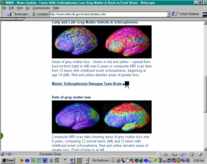
The notation below the pictures indicated the source as:
Paul Thompson, M.D., UCLA, Laboratory of Neuroimaging
If the brain was indeed now known to reorganize right around the onset of puberty, and frontal lobe functions were “coordinated” to a great extent, by the cerebellum – that part of the brain located at the back of the head – that part of the brain also most impacted in autism – would it not stand to reason that the cerebellum - known to be involved in the coordination of frontal lobe functions - would be somehow also involved in the normal reorganization of those areas that should occur right around puberty? And, indeed, if that were the case, what would trigger that “reorganization”? Could it be the cerebellum itself – that part of the brain known to take twenty years to reach maturity – placing the timeframe for the maturity of the cerebellum right around the “end” of the above study in terms of the age of participants at the end of the study! Coincidence?
Again, I was not a scientist, but, when it came to issues such as this, my “coincidence comfort level” had gone out the window a long time ago.
It would make perfect sense that the cerebellum would reach maturity once its “job” had been completed – a job, that in my opinion, very much appeared to be - the reorganization of the brain and processes involving brain structure/function maturation!
As I considered all this – what were the implications? Well, in schizophrenia, I would expect to see the first things impacted to be things like vision – located at the back of the brain, in the occipital lobe. Then, I would expect to see parietal lobe and temporal lobe functions impacted – issues with touch perception, somatosensory processing, auditory processing, memory, emotion, understanding of language, voice and face recognition, ability to distinguish between truth and a lie (the real and non-real), etc. Lastly, frontal lobe functions that included motor functions, language production and higher thoughts (imagination, concept of self, etc.) would be impacted – although, clearly, from the results of the study, one could see some impact to all areas from the very start of the study.
As I looked at these images of the schizophrenia brain, I could not help but wonder if the “early gray matter loss” in the frontal lobe – the damage that appeared to be there even before the study had begun – could be somehow related to the “2 peaks” for schizophrenia when it came to diagnosis. The first peak for diagnosis was around age seven to nine, the second, in boys, was in the early twenties, in girls, in the late twenties. The brain of a girl was known to mature faster and thereby, this perhaps provided more protection against damage, according to Dr. Jay Geidd of the National Institute of Mental Health, per his interview with PBS Frontline – and indeed, also, according to the “Simpsonwood meeting of 2000”. Could this early change in the frontal lobe, captured in the schizophrenia brain experiment explain the “two peaks” in diagnosis – the very early age seven to nine and then the “twenties” peak? In my opinion – it certainly could!
If one considered “what happened” in a child around the age of seven, clearly – perhaps the most obvious change, at least in my opinion, had to do with matters dealing with the “concept of self” and “imagination” – functions located in the frontal lobe. A child’s concept of self certainly did change around age seven, as did his belief “in the tooth fairy”. Children around this age certainly became more aware of not only themselves, but of matters dealing with reality verses imagination. It had also been very well documented that when children entered the school system, they lost a great deal of their “creativity” – a higher function – also in the frontal lobe!
Could these be the area shown as “impacted” very early on in the schizophrenia brain study – even though the general wave of the “impact” was from back to front? Surely, those “red and pink spots” showing gray matter loss in the frontal lobe from very early on could be associated with the concept of self, imagination, and creativity – issues clearly implicated in schizophrenia. In my opinion, there was no denying that these issues, when considered in view of the MRI scans of the schizophrenia brain experiment, clearly could contribute somewhat to the explanation of the “two peaks” in the diagnosis of schizophrenia.
As I thought about this issue of brain development as it related to the development of both white and gray matter, it occurred to me that once the “big wave” of white matter development was completed, perhaps the brain’s metabolism of iron changed somehow. White matter was known to be rich in iron receptors. White matter development seemed to go through tremendous growth between the ages of three and six. By age six, the brain had ninety five percent of its adult size. As such, I could not help but wonder, how did iron metabolism change after age six once that peak in white matter development was initially completed? And, how did this impact iron metabolism – if at all – as it related potentially to the role of iron in the diagnosis of schizophrenia?
In all humans, white matter was believed to develop first and that white matter was enveloped by the gray matter that thickened later in life. White matter received its name from the fact that these cells in the brain were wrapped with myelin – a white protective coating that made neural transmission more effective. Although white matter was associated with earlier brain development, white matter continued to develop through adulthood. So, proportionately, it appeared a young child had more “white matter” than “gray matter” in the first years of development and that “gray matter thickening” occurred during adolescence.
How did this relate to autism – diagnosed so much earlier? In autism, the child had not yet undergone “peak” in white matter development that occurred between the ages of three and six nor had the child undergone the “pre-puberty” gray matter development wave and maturation of the cerebellum – something that took twenty years.
Interestingly, white matter developed in a wave-like fashion too – only in this case the wave of development went from front to back! White matter developed first - from birth on into adulthood and developed myelin sheath to make it more "efficient" over time.
Again, I also wondered about the fact that mercury appeared to have its greatest impact on immature cells. Would mercury target white matter cells because they too, like the cerebellum cells – were immature at birth? Or was white matter targeted more by excess iron given white matter was rich in iron receptors? Or, could both – white matter and the cerebellum - be impacted – at the same time?
Given white matter cells developed in a front to back fashion, would it not make sense that frontal lobe functions – functions that were very much known to be impacted very early on in autism – functions such as language production, concept of self, imagination, motor functions, emotions – would again be the most “hit” and that given myelin had not had the chance to develop properly, that connections within the brain in terms of message relays would be hampered – leading perhaps not only to issues in “transition” due to the “slower connections”, but also to “fragmented thoughts”. If the cerebellum was known to coordinate both motor functions and higher thoughts and the cerebellum was known to reach maturity only after twenty years, it stood to reason that the “coordination of thoughts” would be greatly impacted if the cerebellum, - what in my opinion was the “brains of the brain”- was assaulted in any way prior to reaching maturity.
This certainly would explain why persons with schizophrenia were usually verbal whereas children with autism, primarily, were not. Up to fifty percent of children with autism were considered “non-verbal”!
Autism… schizophrenia… white matter and gray matter development waves – in opposite directions… at different times in life. Needless to say – again – this was all very interesting!
How did all this relate to Alzheimer’s? In Alzheimer’s, clearly, the reorganization, pruning and wave of gray matter development that began around puberty and continued to about age twenty and the maturity of the cerebellum had been completed prior to the onset of the disorder and as such, this had to have implications in terms of what we saw in Alzheimer’s as opposed to autism or schizophrenia.
As I learned more about these disorders and matters relating to development that could explain the differences we saw in autism, schizophrenia, and Alzheimer’s in terms of age, symptoms, etc., I became more and more convinced that, indeed, these were but shades of the same thing – improper neural connections due to an assault on the brain that had led to cell death and hence, resulted in “fragmented thoughts” and overall systematic issues. Clearly, in all three disorders, the entire brain was impacted – and all systems seemed to be impacted. In my heart, I knew this was not a matter of “genetics”.
The brain cell loss was so extensive in schizophrenia, that I was truly, very, very concerned for Zachary. I could not help but ask myself, could mercury be lodging in the area of the cerebellum in young infants, and then, with the puberty gray matter wave of development that occurred in the brain – from back to front – could that wave of development in gray matter, somehow – be taking mercury “along for the ride” - as the wave moved to the front of the brain? Could this have something to do with the tremendous gray matter loss seen in schizophrenia with the onset of puberty? The University Of Calgary video showing neural degeneration due to low- level mercury exposure kept playing through my mind… certainly, if mercury was somehow “flowing along” with the gray matter development wave… this was a possibility – at least in my opinion!
The other possibility was that of iron binding to nitric oxide given nitric oxide synthase seemed most associated with the cerebellum. Heme deficiency, something also related to “iron”, given heme was made of “iron plus unconjugated bilirubin”, was now also known to activate nitric oxide synthase and interfere with iron homeostatis, as stated earlier.
If indeed the cerebellum “triggered” or was in any way involved in that gray matter thickening wave at puberty – if excess iron and/or nitric oxide was found in the cerebellum, and nitric oxide – and iron – in excess – were both known to lead to cell death – as that wave of gray matter development occurred, would iron/nitric oxide levels not - potentially – also be an explanation for the tremendous cell death seen at puberty onset in teens with schizophrenia?
The schizophrenic teenagers involved in the study by the National Institute of Mental Health that showed extensive gray matter loss in schizophrenia were much older than Zachary. These teenagers had been at least fourteen years old and had been followed through age nineteen or so as the study had lasted five years. To me, that clearly meant that these teenagers with schizophrenia had been exposed to much less earlier in life in terms of vaccines – mercury, viruses, etc. - than had been Zachary. These twenty year olds would not have had the extensive and aggressive vaccination schedules Zachary, a child not yet six years old, would have had. If mercury or excess iron or nitric oxide had contributed to this massive devastation in the schizophrenia brain, I had some very serious concerns about what could potentially happen to my son once he reached puberty and his brain began its reorganization and pruning stage. How much gray matter could Zachary lose - a child who had been exposed to much, much higher levels of mercury, and could possibly have excess nitric oxide already forming in the cerebellum. Zachary had also been exposed to more viruses via vaccines much earlier on. What awaited - my son – at puberty onset?
What was causing this loss of gray matter in schizophrenia? I needed some answers – yesterday!
Science had already shown that the neocerebellum (where cerebellum attached to the cerebral cortex) seemed to be "disconnected" in those with autism. Could mercury exposure have caused this? Given the University of Calgary experiment on neural degeneration as a result of mercury exposure, I could not help but wonder.
Was this loss of gray matter in schizophrenia at puberty due to mercury? Or, could this once again be an immune system response to iron overload? Or could this have something to do with viruses in vaccines? In my opinion, it certainly could be any of these possibilities.
Glial cells, found in the brain, appeared to be sensitive to iron and Hans Moises and his team had shown glial cells could be weakened by viruses. Could this loss of gray matter in schizophrenia result from mercury? Mercury had clearly been shown to contribute to neural degeneration by the University of Calgary.
Mercury also seemed to have a propensity for developing cells… it was known to flow from mother to unborn child. Could it be that mercury or viruses first lodges in the area of the cerebellum (affecting the order of thoughts and motions), and then, with the second wave of development and the thickening of gray matter, occurring at adolescence, that the mercury and other metals were then "taken forward" in that growth wave, impacting other areas of the brain? If mercury did have a propensity for developing neural cells – what cells were the most vulnerable?
Hans Moises and his team had also shown that viruses themselves could be implicated in the weakening of glial cells – the cells providing scaffolding in the brain. That certainly could explain why the MMR, a vaccine with no mercury in it, appeared to trigger autism in some children almost overnight. The MMR… a vaccine with three live viruses… viruses… known to thrive on iron! Certainly a “scaffolding” collapse could result in improper neural connections and fragmented thoughts, too.
Nitric oxide… iron overload… viruses… mercury… in my opinion – all excellent possibilities in and of themselves and/or in combination! So many questions… and so very few answers! My search for those answers continued.
Nitric oxide was known to bind to iron… nitric oxide appeared to be very much related to the cerebellum given nitric oxide was synthesized as needed by nitric oxide synthase (NOS)… Nitric oxide synthase (NOS) was found in high concentrations in the cerebellum…
In my opinion, it stood to reason that nitric oxide would be produced in higher concentration where nitric oxide synthase (NOS) was also found in higher concentration – and that would be - in the cerebellum! Nitric oxide was also believed to increase the permeability of the blood brain barrier – and that, certainly, would make the brain more vulnerable to viruses!
Nitric oxide … known to bind to iron… iron overload… found in autism, Alzheimer’s and schizophrenia! Could nitric oxide production have something to do with iron overload? Nitric oxide production level issues had indeed been documented in all three disorders as well. Could this, like high lactoferrin in Alzheimer’s, and abnormal nitric oxide levels be an immune system response in an attempt to rid the body of excess iron? In my opinion, it certainly could be a possibility!
Given that iron appeared to bind to so many things, it was in my opinion, key to know what iron liked to bind to “ the most” – what it most easily bound to and – what it could not be easily “unbound” from and then look at those functions as they related to key organs and the brain over life span development – especially from an immune system response perspective. What did iron bind to the most easily – the most strongly? Was it lactoferrin? Was it this thing called “beta”? Was it nitric oxide? What was the role of mercury or aluminum in iron metabolism? I did not have the chemistry background to understand these issues, but I knew a parent of an autistic child or a scientist – somewhere – would!
I had also found it interesting that a very well known immunologist Dr. Vijendra K. Singh, had two areas of expertise – autism and Alzheimer’s.
In my opinion, given these were supposed to be “separate disorders”, either one of these disorders was a life-long project in and of itself in terms of addressing immune system issues. How was it that this man was an expert in both? Was it because he, too, had seen what I had seen – the many, many parallels between autism and Alzheimer’s? Was it because he was not studying two disorders... but really - only one? Two disorders or, – one?
Was this again, “just coincidence”?
Autism… schizophrenia… Alzheimer’s… mercury… aluminum… viruses… iron… nitric oxide…
I had come to understand a little more in terms of iron than I had first known – now it was time to understand a little more in terms of nitric oxide.
In 1998, Robert F. Furchgott, Louis J. Ignarro and Ferid Murad of the United States had won the Nobel Prize, for the discovery of properties of nitric oxide, a common air pollutant, considered a lifesaver because of its capacity to dilate blood vessels.
I certainly had no doubt that nitric oxide could indeed “dilate blood vessels”, but, that, to me, sounded like something that would be a “symptom” of something going wrong – if nitric oxide “dilated blood vessels”, could that not mean that they were “starving” for oxygen and trying to survive? I was simply a housewife – not a Nobel laureate – but, again, I certainly wondered!
Certainly, the findings of these men were accurate – nitric oxide dilated blood vessels – of that there was no question. What I questioned was not their discovery. What I did question, however, was how we came to view nitric oxide as a neurotransmitter – something that would seem to imply “it belonged” in the brain, when clearly, so much literature showed the very detrimental effects of nitric oxide on human cells. Nitric oxide was very much associated with “cell death” and that alone, I found to be very, very troubling – especially given that nitric oxide synthase appeared to be in high levels in the cerebellum – the very part of the brain impacted in autism!
There were many substances now found in the human body – but – did that mean they necessarily belonged there? That was “the” question – at least in my opinion.
What exactly was a neurotransmitter? I typed that into The Britannica Concise online encyclopedia and obtained the following:
“Chemical released by neurons to stimulate neighboring neurons, allowing impulses to be passed from one cell to the next throughout the nervous system. A nerve impulse arriving at the axon terminal of one neuron stimulates release of a neurotransmitter, which crosses the microscopic gap (see synapse) in milliseconds to the adjoining neuron’s dendrite. Many chemicals are believed to act as neurotransmitters. The few that have been identified include acetylcholine, dopamine, and serotonin. Some neurotransmitters activate neurons; others inhibit them. Some mind-altering drugs act by changing synaptic activity” [end of quote, emphasis added: The Britannica Concise online encyclopedia available at: http://education.yahoo.com/search/be?lb=t&p=url%3An/neurotransmitter].
Note that in defining a neurotransmitter, according to The Britannica Concise online encyclopedia, there was no distinction between a “good”, or a “bad” neurotransmitter. A neurotransmitter, according to The Britannica Concise online encyclopedia was simply something that was chemical in nature – mercury, aluminum, iron and nitric oxide were all “chemical in nature” - and provided a stimulant for neurons to engage in “reactions”.
Thus, science was correct in this regard – nitric oxide was a neurotransmitter – but, was it a “good one” - was it a neurotransmitter that belonged in the body? In my opinion, there could be no doubt that certain substances could “impact” transmissions within the brain, but whether all those “things” were necessarily “good” was another matter.
There were now literally thousands of websites dealing with the subject of nitric oxide and there appeared to be simply too many indicating the huge “negatives” associated with abnormal nitric oxide production. But, what did we “know” about nitric oxide?
Although the Nobel Prize had been won for the properties of nitric oxide as they related to the blood, science had discovered a great deal more when it came to nitric oxide. Indeed scientists Brown, G. C., Bolanos, J. P., Heales, S. J. R. & Clark, J.B. (1995) Neuroscience Lett. 193, 201-204, found that nitric oxide inhibits cellular respiration causing a release of glutamate which can potentially be neurotoxic. McNaught, K. St. P. & Brown, G. C. (1998) J. Neurochem. 70, 1541-1546 made similar findings.
Eric Courchesne had scientifically proven the cerebellum of children with autism was much smaller than the cerebellum of "normal" populations. Could that due, not to “genetics” but to cell death due to mercury, aluminum, or iron toxicity possibly resulting in excess nitric oxide? Could this be why we saw so much gray matter cell death in schizophrenia, in Alzheimer’s – in any neurodegenerative disorder?
For those of you in science, Peter Lipton of the Department of Physiology, University of Wisconsin School of Medicine had written an article entitled: Ischemic Cell Death In Brain Neurons that appeared rather interesting. This article was a 138-page review of over 1300 scientific articles on this subject of cell death, published in Physiological Reviews, Vol. 79, No. 4, in October of 1999. The text of this review was available on the following website: http://ntp.neuroscience.wisc.edu/faculty/fac-art/Lipton79.pdf.
I did not have the necessary background to even begin to understand this article, but I wanted to provide it as a reference for those parents of children with autism who did have a good scientific background and were interested more in reading about issues relating specifically to cell death, because surely, within these pages, there had to be more clues to Breaking The Code: Putting Pieces In Place!
Although nitric oxide was associated with cell death, it was also associated with many other functions. It was known to play a role in memory formation, in respiratory disease and pulmonary circulation in newborns (persistent pulmonary hypertension), brain development, pain physiology, diabetes/insulin levels, immune system functions, vision, regulation of enzymes, mitochondria functioning, gastrointestinal track repair, synapse efficacy in the brain, control of blood flow, impact on “other neurotransmitters” (i.e., serotonin, norepinepherine, dopamine, etc), a possible role in MS and demyelination of brain cells, bone and joint disease – also considered immune system issues, etc.
As I looked at this list, there was simply no denying that nitric oxide impacted “things” known to be problem areas in autism, schizophrenia and Alzheimer’s – as well as I was sure, many, many other disorders, such as diabetes, ALD, Parkinson’s (also considered a disorder with iron overload – a sign of mercury toxicity was shaking of the hands), cancer (immune system) and so much more.
Although science wanted us to believe that nitric oxide was a “neurotransmitter” – and I had no doubt that it did impact neural transmissions – I could not help but ask, was presence of nitric oxide in the human body a “good” or a “bad” thing? Did nitric oxide really belong in the human body? Nicotine, alcohol, marijuana, medications – all these impacted neural transmissions too – but, obviously – they did not belong in the body.
Would this matter of nitric oxide be another one of those things that science would later “disprove”? I just had that “really hard time” issue with the fact that a pollutant and known toxic gas outside the body could be “good” within it – especially given nitric oxide’s clear association with cell death and so many things that just were known to “go wrong” in autism, schizophrenia and Alzheimer’s – and indeed, so many other “disorders” known to man!
I just had a very, very hard time reconciling the fact that something that could apparently do so much damage in the body – actually belonged there! I knew iron was definitely needed in the body… and I now knew that iron, although necessary to so many functions, could certainly do tremendous damage in excessive amounts. Perhaps the issue was thus simply one of “levels” or “how much” was present in the body. I just had to know more about “nitric oxide”!
Back to The Britannica Concise online encyclopedia I went in search of more information on “nitric oxide”! Certainly all parents of children with autism, like me, would recognize the last word in the first sentence – “mercury”! I quote:
“Nitric oxide: Colorless, toxic gas (NO), formed from nitrogen and oxygen by the action of electric sparks or high temperatures or, more conveniently, by the action of dilute nitric acid on copper or mercury. First prepared c.1620 by J. van Helmont, it was first studied in 1772 by J. Priestley, who called it "nitrous air." An industrial procedure for the manufacture of hydroxylamine is based on the reaction of nitric oxide with hydrogen in the presence of a catalyst. The formation of nitric oxide from nitric acid and mercury is applied in a volumetric method of analysis for nitric acid or its salts. [emphasis added - end of quote from The Britannica Concise online encyclopedia, available online at: http://education.yahoo.com/search/be?lb=t&p=url%3An/nitric_oxide.]
Well, needless to say, I was certainly not a biochemist… and, although I did not understand the implications of this, seeing the words “nitric oxide” and “mercury” in the same sentence concerned me greatly since I knew that nitric oxide synthase appeared to be in rather high concentrations in the cerebellum – the very part of the brain that appeared most impacted in autism! I knew I did not have the chemistry background to figure this out, but – if there could be a connection between nitric oxide and mercury – in the human body – and nitric oxide was a known toxic gas, then, let us simply say that I had “concerns” that had now been magnified.
The gray matter loss seen in schizophrenia from the onset of puberty weighed heavily on my mind – teenagers with schizophrenia had been shown to have four times the normal loss of gray matter!
My son – my beautiful son – had been exposed to so much more mercury than the participants in that study. More mercury… in a much more compressed timeframe given that over time, vaccination schedules had become more and more aggressive, including not only more mercury, but more vaccines – more viruses – more for the immune system to deal with!
When I had first read this definition of nitric oxide - I had only really noticed that word – mercury! But then, as I read the paragraph a second – and, a third time – again, my heart melted within me… before my eyes, more reason for concern…
Nitric oxide –according to The Britannica Concise online encyclopedia, available at: http://education.yahoo.com/search/be?lb=t&p=url%3An/nitric_oxide, was defined as:
Nitric oxide: Colorless, toxic gas (NO), formed from nitrogen and oxygen by the action of electric sparks or high temperatures or, more conveniently, by the action of dilute nitric acid on copper or mercury. First prepared c.1620 by J. van Helmont, it was first studied in 1772 by J. Priestley, who called it "nitrous air." An industrial procedure for the manufacture of hydroxylamine is based on the reaction of nitric oxide with hydrogen in the presence of a catalyst. The formation of nitric oxide from nitric acid and mercury is applied in a volumetric method of analysis for nitric acid or its salts. [emphasis added - end of quote from The Britannica Concise online encyclopedia].
“That part” about the formation of nitric oxide from “electric sparks” now also jumped out at me given that the brain was very much considered an “electric type” organ when it came to “neural transmissions”. I knew that children with autism, and persons with schizophrenia or Alzheimer’s so often developed seizures (electric impulses) and needless to say, that also put me very much in the “suspicious of all this” mode when it came to nitric oxide in the human body!
I read the paragraph again – another reason for possible concern – that “copper part” of the statement. I knew that zinc-copper metabolism was also impacted in autism, schizophrenia and Alzheimer’s! And, finally, another reason for concern… “high temperatures”…
Children who were vaccinated often had fevers just following vaccinations – would these fevers be high enough to produce nitric oxide? I did not know, but needless to say, like iron, mercury and aluminum, nitric oxide had very much become a key area of investigation.
Nitric oxide…known to bind to iron… nitric oxide… associated with cell death… mercury… electric sparks…copper…seizures… the cerebellum… nitric oxide synthase… nitric oxide… neurotransmitter…cell death… cell death… cell death…
Although I was not a scientist able to understand all of the chemical reactions and what all this meant from a scientific perspective – and as much as I hoped none of this was “related” to my son’s autism – that none of this had anything to do with what was going on in these disorders – I could not help but ask again – was all this “just coincidence”?
My heart once again experienced that recurring “stabbing feeling” that had now become all too familiar throughout this journey with “autism”. How long would there be denial of at least a “possible link”? This was, yet again, “just my theory – a theory”, and now more than ever, a theory I hoped would be proven inaccurate. I truly hoped I was wrong in all this… but my heart – the melting heart of a mother - was telling me otherwise as I thought about how much of my son’s brain, too, could, at the onset of puberty, simply – melt away.
My beautiful son… my only son… exposed to so much mercury via vaccines and possibly also via my dental amalgams… and perhaps, exposed to excess levels of iron during pregnancy via prenatal supplements. I had worked so hard to remove from my son the shackles of autism, and yet, with the onset of puberty and the gray matter loss I very much suspected would occur in my son at that time, more than ever, I felt I could lose him – again! There were truly no words to describe how painful this journey with “autism” had become! More than ever - I felt the clock “ticking” - as I continued to search for answers to my son’s autism.
The cerebellum, I soon discovered, was closely associated with the basal ganglia – another part of the brain impacted in autism – and that part of the brain now believed to be the body’s “clock mechanism”. How interesting that a part of the brain, known to be the body’s “clock” was so closely associated with the cerebellum – a major “coordinator” of so much in the human brain!
The basal ganglia was involved in functions relating the regulation of movement and the learning of skills. This part of the brain also controlled the intensity of mental activity.
If you think about it, a child at birth had a very different schedule in terms of "life functions" than say a five year old, or someone who was older still. For example, babies were always said to just "eat, poop and sleep". By age five, the need to sleep during the afternoon was usually completely gone. Toilet training also occurs well after birth, as did changes in digestive processes (i.e., the liver began to produce bile at age six months, etc.).
So, both the liver and the basal ganglia now appeared to have “clock mechanisms” of some kind.
Not only was this part of the brain implicated in autism, as confirmed by Dr. Eric Courchesne, but it was also very much implicated in Alzheimer’s as well. Indeed, in Alzheimer’s, the basal ganglia had been shown to be high in iron.
In his interview with PBS Frontline: Inside The Teenage Brain, Dr. Jay Giedd of the NIMH also mentioned that girls had a larger basal ganglia, believed to assist in frontal lobe executive functions and as such, he had stated that girls may be "better protected" in terms of frontal lobe damage. The brain of girls, also according to Dr. Jay Giedd, was also known to "mature faster" and as such, was offered more protection than the male brain since the male brain remained "immature" longer.
It was a well-known fact that more boys than girls had autism... with almost four boys impacted for every one girl. The pieces of the puzzle certainly appeared to be falling into place!
Given comments made at the Simpsonwood meeting of 2000 on mercury, and statements by participants of that meeting whereby they admitted that they knew the earlier the exposure to mercury, the worse the effect – there certainly appeared to be cause to believe that the more immature the cells, the greater the potential damage. Thus, this also, when combined with Dr. Jay Geidd’s observations as they related to the basal ganglia and the frontal lobe and differences in the brain “maturation process” between boys and girls, would be very much in line with the fact that more boys were impacted by autism.
The basal ganglia was involved in motor functions and memory. The basal ganglia was involved in the "planning and execution" of motor activities and as such, those with basal ganglia damage would have a difficult time in this area as well as in "sequencing tasks". Some scientists also appeared to believe that when two motor functions needed to be performed - one subconscious and one conscious - for example, that the conscious one would "win out" and be completed. [R.S. Schwab, M.E. Chaefetz, and S. Walker. Control of two simultaneous voluntary motor acts in normals and parkinsonism. Archives of Neurology and Psychiatry, 72:591--598, 1954, referenced online along with other very interesting materials on the basal ganglia at http://www.hitl.washington.edu/publications/prothero/node54.html].
That again was very, very interesting given what I had seen in my son in matters relating to the conscious and subconscious.
This was very interesting given that so many children with autism were unable to complete the proper "unconscious" or instinctive movement necessary to get out of a dangerous situation (i.e., to get away from an oncoming car).
The basal ganglia – involved in the processing of conscious and unconscious tasks… closely associated with the cerebellum… involved in the tracking of moving objects and the thalamus… known to be involved in matters of “consciousness” and the integration of sensory information between the central nervous system and the peripheral nervous system!
The basal ganglia – known to have elevated levels of iron in Alzheimer’s! I quote:
The new imaging technique, known as FDRI (Field-Dependent R2 Increase) measures the iron content of ferritin in the brain. Ferritin is a protein that stores most of the body's iron reserves. The measurement is made through the combined use of two MRI machines with different imaging strengths, says Bartzokis, who is affiliated with the departments of psychiatry at both UCLA and the University of Arkansas for Medical Sciences in Little Rock. Thirty-one people with mild to severe Alzheimer's disease and 68 people who served as controls participated in the study. Ferritin iron levels in different regions of the brain were compared in the two groups. In the Alzheimer's group, ferritin iron levels were significantly higher than levels in the control group -- but only in the basal ganglia, areas traditionally believed to be implicated in movement. [end of quote, emphasis added: Jeanie Lerche Davis , High Iron Levels Identified in Brains of Alzheimer's Patients, WebMD Medical News Archive, Feb 28, 2000, http://my.webmd.com/content/article/21/1728_55296]
Zachary’s difficulty with “seeing moving cars” – was this the reason? It certainly appeared to be a very strong possibility! Indeed, a squirrel appeared to have much more fear of an oncoming car than did my son.
Note that - as in the case of the cerebellum - the functions of the basal ganglia very much involved learned functions (i.e., task completion issues)! As such, once again, we were seeing an area of the brain that was impacted in autism as well as in schizophrenia and Alzheimer’s - and that area seemed to deal primarily with "learned motor functions and memory for completing those functions". Given the role of "learning" being involved in this particular part of the brain, again supported the theory that the basal ganglia most likely had very immature cells at birth... and that given mercury appeared to target or impact "developing cells" more harshly – based on what was learned from the “Simpsonwood meeting of 2000 on mercury” - this would help explain, at least in part, why the basal ganglia was yet another area so impacted in those with autism, iron or mercury poisoning. And, again, this seemed to argue against the "genetic" theory to these disorders since those areas most impacted were areas of the brain that dealt with learned activities - activities that were very much influenced by one’s environment.
Damage to the basal ganglia area certainly helped to explain why potty training was so difficult for so many of these children. Indeed, science had by now determined that the basal ganglia actually received information (input) primarily from the motor cortex and somatosensory cortex - and that, appeared to implicate regions involved in "potty training".
Visual attention, goal directed movements, and integration of sensory input to allow for the understanding of a single concept – like danger – all functions in the parietal lobe – where the somatosensory cortex was located!
Output from the basal ganglia, however, seemed to go to the motor cortex and the brain stem - where life functions resided - and those "life functions" included reflexes to sight/sound and alertness levels. This certainly seemed to explain – so much!
When cars – or bulls – as mentioned in my second book – were stationary – Zachary could “see them”, yet, when they became “moving objects” – dangerous moving objects – as Zachary focused on his “real world” all about him – did he fail to do the subconscious task of “danger assessment” and fail to move out of the path of dangerous oncoming objects! This processing of the conscious taking precedence over the subconscious – again – simply appeared to explain – so much!
As such, again, certainly, it was very likely that the basal ganglia was involved in matters of "potty training" and in matters of "fight or flight" - or reflexes to one's environment and the production of the appropriate response to that environment (i.e., an oncoming car). These were "learned responses" that became "automatic"... until you were "potty trained" and taught to "move if a car was coming", that response would not be there! Learned responses… that then became “automatic. The conscious, that then became almost “subconscious”.
Science also seemed to indicate that basal ganglia output was inhibitory in nature, whereas that of the cerebellum was "excitatory" in nature. I did not understand all the implications of this, but wanted to mention this for parents who would understand what this meant.
The role of the basal ganglia, in the last twenty years, had come to be much better understood. We now knew the basal ganglia to be involved in all types of goal directed behavior. The basal ganglia were believed to play a role in much more than just "motor functions". It was now believed the basal ganglia played a role in those things that drive behavior - emotions, motivation, rewards, and other cognitive processes. As such, the basal ganglia were believed to play a role in obsessive-compulsive behavior, addictive behaviors, habit formation, and working and/or procedural memory as well (i.e., in learning to ride a bike). Could this possibly have anything to do with why conscious activities, such as gambling, became obsessive and “almost subconscious” over time? Very interesting indeed!
The basal ganglia also appeared to play a role in "mastication" - motor functions involved in eating/chewing your food. Neural connections did indeed flow from the basal ganglia to the brainstem - involved in swallowing and digestive processes. As such, damage to the basal ganglia certainly could play a role in "swallowing" issues – something so very common in autism and Alzheimer’s.
I now understood a little more about the basal ganglia and its functions… but, most important perhaps, was the realization that in Alzheimer’s, the basal ganglia appeared to be high in iron! There was that word again – iron!
Iron… the brain… changes of time… in metabolism… in development… Clearly, there was no doubt that to properly understand these disorders, you had to look at all these issues. My search for answers to my son’s autism continued…I looked to understand more in terms of brain development and how it could explain the differences seen in these three disorders – autism, Alzheimer’s and schizophrenia.
The PBS Frontline interview with Dr. Jay Geidd entitled Inside The Teenage Brain had provided for me so many insights as they related to matters involving the cerebellum. As I had done on so many occasions before, I went back and took “a second look” at what had been provided by Dr. Jay Geidd of the National Institute of Mental Health. More pieces to the puzzle…
In this interview, Dr. Jay Geidd had also mentioned that the corpus callosum was undergoing great changes during this stage – adolescence - also. The corpus callosum was a collection of fibers that connected the left and right hemispheres of the brain. This part of the brain allowed the left and right hemispheres to “speak” to or communicate with one another.
The corpus callosum was now also known to undergo great growth before age fifteen. As such, this was another "immature area" that would be susceptible to mercury if mercury indeed did target or have a greater effect on “developing or immature cells”.
In an article that appeared in the LA Times-Washington Post, entitled: "Study reveals how human brains grow" by Robert Lee Hotz on the work of UCLA neurologists Arthur W. Toga and Paul Thompson amazing pieces of the puzzle may have been revealed. These neurologists found that between ages three to six, most growth in the brain was in areas for the learning of new skills (i.e., learning to think ahead, attention focus, alertness, planning of new actions). Most of the growth seen was in the frontal cortex and moved from "front to back".
This certainly was very interesting given this was the very time at which autism was usually diagnosed. There was no doubt in my mind that the cerebellum and basal ganglia had been impacted in autism – that had clearly been shown. Many parents were pointing the finger to vaccines, others, to different factors. In my opinion, it was a combination of several factors that appeared to lead to autism – factors that included mercury and aluminum poisoning, nitric oxide production excesses, iron overload and potentially, viruses themselves.
Science had established that the brain reached approximately ninety-five percent of its adult size by age six. As such, growth in the brain, prior to age six, had to hold critical pieces to the autism puzzle. I now began to consider what happened in the brain during this timeframe… age three to six. Clearly, the areas mentioned for period of development – learning to think ahead, attention focus, alertness, planning new actions – involved the cerebellum and basal ganglia – parts of the brain known to be very much implicated in frontal lobe and parietal lobe functions.
Ages three to six… early brain development… front to back… iron overload…nitric oxide production…mercury… early brain development…white matter… associated with earlier development and “child-like behaviors”… white matter formed first... in the earlier years of life...and continued to form through adulthood… white matter… “child-like behavior”… “regression” to child-like behaviors” in autism, schizophrenia, and Alzheimer’s… myelin formation… neural efficacy…iron… nitric oxide… mercury…viruses…
White matter… gray matter…
“Gray matter" formed last, and somehow "enveloped" the white matter... in other words, the "gray matter" was "on the outside", with the "white matter" on the inside. If gray matter was on the “outside”, what effect would that have, if any, when exposed to neurotoxins such as mercury, aluminum or excess iron and/or nitric oxide? Given the University of Calgary video, there could be no doubt that mercury had a tremendously negative impact on neurons – but, what neurons – gray or white – or both?
Mercury and aluminum would have to cross the blood brain barrier to get to the brain. Nitric oxide, something known to bind to iron, was known to increase the permeability of the blood brain barrier. Nitric oxide was produced by “electric sparks” (the brain had electric currents, and those with autism, schizophrenia and Alzheimer’s developed seizures), high temperatures (fevers?), or as a result of dilute nitric acid on copper or mercury.
The blood brain barrier was immature at birth and not fully formed until at least six months. If mercury crossed the blood brain barrier and made it to the surfaces of the brain, there could be no doubt, based on the University of Calgary experiment on neural degeneration due to low level mercury exposure, that neurons would absolutely be impacted. Thus, if crossing the blood brain barrier, mercury would – it would seem – come in contact first with “gray matter” and hence, it would make sense that mercury would have – at least initially – an impact on the gray matter.
If gray matter was destroyed, would that not leave “white matter” to be found in a proportionately greater amount and also, perhaps leave the “white matter” – physically – more exposed and hence – potentially – more susceptible to mercury exposure also, thereby, leading to destruction - potentially – of white matter also. Would a loss in gray matter not help explain “regression” in behavior to more “child-like” behaviors if gray matter – associated with “maturity” were somehow destroyed and “white matter” – associated more with child-like behaviors and early development – were more exposed or found in greater proportion due to the loss of gray matter? There was no doubt that mercury, certainly could play a role – but what else could lead to such devastation in the brain? Was white matter impacted first – gray matter, or, - were they both being “hit” at the same time?
White matter or, - gray, or - both? In my opinion, it looked like the issue was one of “both” being destroyed. White matter was clearly associated with the efficacy of neurons given that myelin made neural transmissions “more effective”. Persons with autism, schizophrenia and Alzheimer’s certainly had issues with “transitions” from one thought to another, from one activity to another, etc., and that, appeared to indicate problems in communication among the various parts of the brain – problems with “neural transmissions” leading to ineffective transitions. Problems with “transitions” or improper connections could very much be due to mercury exposure or the action of iron on white matter cells, known to be rich in iron receptors.
Mercury had been shown to completely devastate neurons – literally “frying them” and reducing them, significantly, in size with even low-level exposure. Neurons exposed to mercury were not only “fried” by the mercury, they were also very much impacted in terms of future growth also, and seemed to go on to form neurofibrillary tangles – all things that had been found to be true by the University of Calgary experiment. There simply could be no denying that mercury, if it crossed the blood brain barrier, could absolutely devastate neurons!
Given that the brain had “folds”, would mercury seep into those folds – perhaps having more access to critical white matter and limbic system structures such as the amygdale (emotions), hippocampus (memory) and so on – structures all known to be clearly impacted in autism, schizophrenia and Alzheimer’s also. It certainly could be possible – could it not?
Mercury – there could be no doubt – was absolutely a possibility when it came to the destruction of gray and/or white matter – especially given what had been said in the Simpsonwood meeting of 2000 and what had been shown by the University of Calgary experiment!
Although most had associated Alzheimer’s issues in “gray matter”, research conducted by Alex Roher, M.D., Ph.D., director of the Sun Health Research Institute in Sun City, Arizona (http://www.eurekalert.org/pub_releases/2002-09/acs-adm091802.php), was now indicating that perhaps it was the white matter that was first impacted in Alzheimer's - causing demylenation of nerve cells! Dr. Roher’s research findings were originally published in a Sept. 17 print edition of Biochemistry. This was a peer-reviewed journal of the American Chemical Society.
Although mercury was certainly a likely contributor to neural degeneration in these disorders – what about iron and iron overload? Iron also accumulated in the brain – and so much now seemed to indicate that iron overload, too, could be a problem. Iron rich receptors were known to be located in the white matter of the brain.
Furthermore, research done by LD Wedewer, US Autism Ambassador and Kathy Blanco, mother of two children with autism, certainly indicated that iron overload was very much associated with myelin issues (white matter) and matters involving the proper functioning of glial cells – cells that provided the “scaffolding” for the brain!
High lactoferrin levels had been found in the spinal fluid of persons with Alzheimer’s. Iron could bind to nitric oxide. Nitric oxide was definitely associated with cells death when present in excessive amounts. Nitric oxide production was associated with “electric sparks, high temperatures” or could be produced as a result of dilute nitric acid on copper or mercury.
High iron levels had also been found in the basal ganglia of persons with Alzheimer’s. Heme (iron plus unconjugated bilirubin) deficiency had been associated with the activation of nitric oxide synthase (NOS), altered amyloid proteins, the inhibition of zinc and iron homeostasis as previously mentioned.
So many issues to consider… so many bits and pieces to put together…